How aware are you? – Brain Aneurysm Awareness Quiz
The Brain Aneurysm Foundation invites you to challenge yourself and spread awareness. Start by reading this article then clicking on the quiz at the bottom to test your knowledge. Re-take it as many times as you’d like and don’t forget to share your results with family and friends! Good luck!
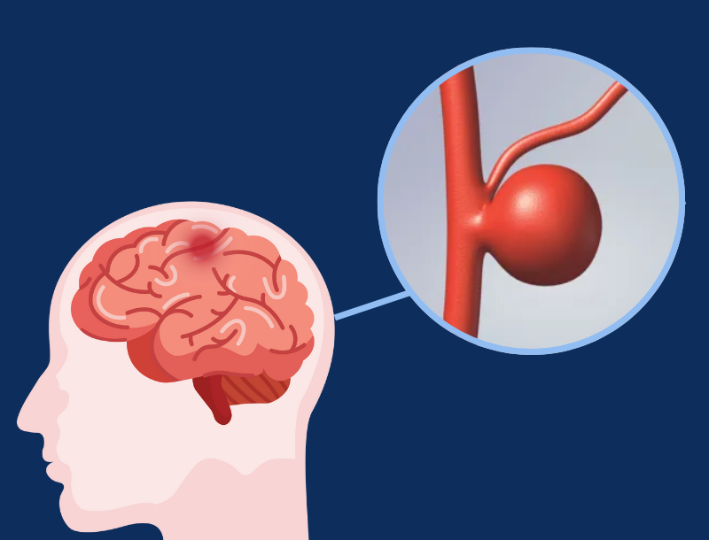
A brain aneurysm, also called an intracranial, cerebral or berry aneurysm, forms because an artery in the brain has a weak spot. Over time, the blood pumping through brain blood vessels causes this weak and vulnerable spot to balloon or bulge. This “ballooning” can form aneurysms of all sizes from small to large. Most are small (about 1/8 inch to one inch) but can have devastating and life-long consequences if they rupture. This rupture is much like a balloon popping.
If an aneurysm ruptures, blood spills into the subarachnoid space located between the skull and brain putting pressure on the brain. This bleeding can also cause a serious type of stroke known as a subarachnoid hemorrhage. In fact, 3-5% of all new strokes are caused by a brain aneurysm rupture.
There’s about a 50% chance of surviving a rupture and of those who do, 66% suffer a permanent neurological deficit. Seeking medical attention immediately is key to having the best outcome.
Common symptoms of a rupture are:
- A sudden severe headache often described as the worst headache of your life
- Nausea/vomiting
- Stiff neck
- Blurred or double vision
- Sensitivity to light
- Seizure
- A dilated pupil
- Pain above and behind the eye
- Loss of consciousness
- Confusion
- Weakness and/or numbness
- Drooping eyelid
Brain aneurysms develop silently forming in approximately 6.8 million people in the United States, which is equivalent to 1 in 50 people. Much about brain aneurysm formation is still unknown but most of them probably develop as a result of wear and tear on the arteries throughout a person’s lifetime or due to an injury to the head. In some cases, people may have inherited a tendency for weak blood vessels. In return, this could result in an abnormality of the artery wall that will cause the aneurysm to form.
Individuals in families where there are two or more first-degree relatives (parent, child or sibling) with proven aneurysms are at higher risk. These are called familial aneurysms and these family members should go through routine screenings.
Additional risk factors that contribute to their formation are:
- Smoking
- Excessive alcohol use
- Drug use, particularly cocaine.
- High blood pressure (hypertension)
- Age – over 40
- Gender – Women are more likely than men to have a brain aneurysm at a 3:2 ratio
- Race – African Americans and Hispanics are twice as likely to have an aneurysm than Caucasians
- Autosomal polycystic kidney disease

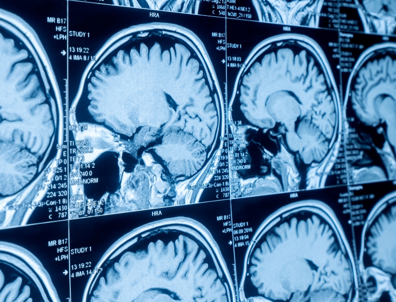
Did you know every 18 minutes a brain aneurysm ruptures? Here are four imaging tests used to diagnose aneurysms before and after a rupture.
Computerized Tomography (CT) or Computerized Tomography Angiography (CTA)
The most common procedure used to detect a brain aneurysm. A CT scan will show if there has been bleeding in the brain; however, it does not show the cause of it. To get a more detailed look at the blood flow in the brain’s arteries, a computerized tomography angiography (CTA) may be ordered instead. This process involves injecting a contrast dye into the bloodstream to highlight the brain’s blood vessels.
Magnetic Resonance Imaging (MRI)
A non-invasive procedure that allows physicians to see a detailed image of the brain by using computer-generated radio waves and a powerful magnetic field. This procedure is commonly used to look for unruptured brain aneurysms.
Magnetic Resonance Angiography (MRA)
A procedure that allows physicians to see a detailed image of a patient’s blood vessels and detect structural abnormalities. This also uses computer-generated radio waves and a powerful magnetic field but can be done with or without dye.
Angiogram
Takes a closer look at the arteries in the brain providing a better view of an aneurysm or vascular malformation. The radiologist passes a catheter up from an artery in the groin to the arteries in the neck; he or she then injects dye into the carotid and vertebral arteries while multiple x-rays are taken of the arteries in the brain.
Help us spread awareness and advocate for brain aneurysm funding so patients and doctors get the knowledge they need to find a brain aneurysm before it ruptures!
Start by sharing this article and knowledge quiz. Many people are unaware that they have a brain aneurysm, routine screenings are not prescribed, and, in many cases, symptoms caused by a brain aneurysm are often misdiagnosed as other problems such as migraines, stress or anxiety.